News & Events
Understanding Size Exclusion Chromatography in Protein Research

Sep

You use size exclusion chromatography to separate molecules by size, making it a key method in protein analysis. This technique helps you work with proteins, peptides, viral vectors, nucleic acids, and lipid nanoparticles. You can determine molecular size, but you need to calibrate your system carefully because accuracy depends on this step. Other methods, like multi-angle laser light scattering, can give you direct measurements, but SEC remains popular for its simplicity. Following best practices helps you get reliable results every time.
Key Takeaways
- Size exclusion chromatography (SEC) separates molecules based on size, allowing for the analysis of proteins, peptides, and nanoparticles without altering their integrity.
- Choosing the right gel beads and pore sizes is crucial for effective separation; match the pore size to your target molecule for optimal results.
- Follow best practices in sample preparation and column care to ensure accurate results and extend the life of your SEC column.
- Use calibration curves to estimate molecular sizes accurately; this step is vital for reliable protein analysis and quality control.
- SEC is essential for purifying proteins and detecting aggregates, making it a key technique in biopharmaceutical development and quality assurance.
Size Exclusion Chromatography Basics
Principle of Separation
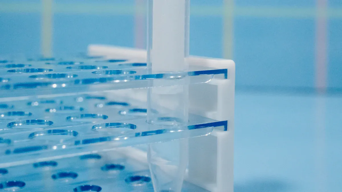
You use size exclusion chromatography to separate molecules based on their size and shape. In SEC, you load your sample into a column packed with tiny gel beads. These beads have pores of different sizes. When you run your sample through the column, larger molecules move around the beads because they cannot enter the small pores. Smaller molecules slip into the pores and take a longer path. This means larger molecules come out of the column first, while smaller ones elute later.
Tip: SEC does not rely on chemical interactions. You preserve the integrity of your proteins and other molecules during separation.
Here is how the process works:
- Larger molecules elute faster than smaller ones.
- The separation depends on the hydrodynamic volume, which relates to the Stokes radius of each molecule.
- You do not need to worry about chemical reactions inside the column.
- SEC helps you keep your sample pure and intact, which is important for protein analysis.
Gel Beads and Pore Sizes
The columns in SEC contain gel beads with controlled pore sizes. You choose the right column based on the size range of the molecules you want to separate. The beads are usually made from cross-linked polymers, such as polystyrene or silica. These materials give the beads strength and stability.
- Common materials for gel beads include cross-linked polystyrene, silica, and other polymers.
- You select the material based on your sample and the solvent you use.
- The pore size in the beads determines which molecules can enter and which cannot.
If you use beads with smaller pores, you separate smaller molecules with better resolution. Larger pores help you separate bigger molecules. For example, Superdex 200 Increase columns let you separate proteins from 10,000 to 600,000 Daltons. This range is perfect for antibodies between 100,000 and 300,000 Daltons. Silica-based SEC columns offer higher pore volume, which improves resolution and selectivity for large molecules.
| Bead Material | Typical Use Case | Pore Size Range |
|---|---|---|
| Cross-linked Polystyrene | General protein separation | 10,000 – 600,000 Da |
| Silica | Large protein and nanoparticle analysis | Up to 2,000,000 Da |
| Other Polymers | Custom applications | Variable |
Note: A narrow distribution of pore sizes in the beads gives you sharper separation and better results.
You need to match the pore size to your target molecules. If you want to separate small peptides, choose beads with small pores. For large proteins or nanoparticles, pick beads with larger pores. The mechanical strength of the beads also affects how well your SEC column works. Strong beads keep their shape and help you get consistent results.
SEC Process Steps
You follow a clear workflow when you use size exclusion chromatography. Each step helps you get accurate and reliable results.
- Column Preparation: You start by packing the column with porous beads and equilibrating it with buffer. This step sets up the right conditions for separation.
- Sample Injection: You inject your prepared sample into the column. Use a sample loop or injection port for this step.
- Elution: The buffer, called the mobile phase, carries your sample through the column. Larger molecules move around the beads and come out first. Smaller molecules enter the pores and take longer to elute.
- Detection: As molecules leave the column, a detector measures their concentration. You see this as peaks on a chromatogram.
- Data Analysis: You use the retention times or volumes to estimate molecular size and check purity.
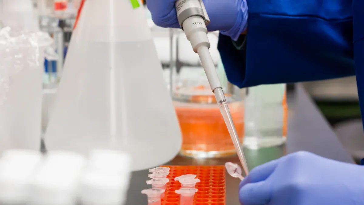
Sample Loading
Proper sample loading helps you get sharp, reproducible peaks. Here are some best practices:
| Best Practice | Description |
|---|---|
| Managing Sample Load | Do not overload the column. Follow the manufacturer’s load limits to avoid distorted peaks. |
| Sample Preparation | Filter your sample with a 0.22 µm filter. Keep the concentration within recommended limits to prevent peak broadening. |
| Mobile Phase Conditions | Use a buffer that prevents aggregation and has low viscosity. Degas the buffer to avoid bubbles. |
Tip: Clean and filtered samples protect your column and improve your results.
Elution and Detection
You choose a detector based on your sample and analysis needs. Here are common detector types:
| Detector Type | Description |
|---|---|
| UV Detectors | Easy to use, sensitive, and good for proteins at 280 nm. |
| Mass Spectrometry | Precise for small molecules, but complex samples may be challenging. |
| Light Scattering | Sensitive to large molecules and useful for samples with many sizes. |
| Refractive Index | Often used with light scattering; needs extra information for accuracy. |
| Fluorescence | Very sensitive for certain proteins. |
| Viscometers | Detect changes in structure and help spot aggregation or denaturation. |
You monitor the elution profile to see when each molecule leaves the column. This helps you identify and quantify your sample components.
Calibration Curves
You use calibration curves to estimate the molecular size of your sample. Here is how you build them:
- Plot the logarithm of molecular mass (log(MW)) against retention time or volume.
- Use standards like polystyrene or monodisperse polymers for calibration.
- Sometimes, you use broad standards or universal calibration to cover more types of molecules.
- Measure at least 10 standards to make a reliable curve. The curve is often non-linear and depends on your column.
Accurate molecular weight determination can be tricky. Using the right standards for your sample type improves accuracy. Some researchers use conversion equations to get better results, especially for complex polymers.
Applications in Protein and Biotherapeutics
Protein Purification
You often use size exclusion chromatography when you need to purify proteins for research or production. This method separates proteins by their size, so you can collect the exact protein you want. You do not need to rely on chemical interactions, which helps you keep your proteins in their natural state. This is important for protein analysis because you want to study proteins as they exist in real life.
When you work with biopharmaceutical products, you need to remove unwanted forms, like aggregates or fragments. Size exclusion chromatography helps you do this by letting you collect only the pure protein. You can also use this method to separate viral vectors and lipid nanoparticles from other components. This makes it easier to prepare samples for further testing or for use in therapies.
Tip: Always match your column pore size to your target protein or nanoparticle. This helps you get the best separation and highest purity.
Aggregation Analysis
Protein aggregation can cause problems in both research and medicine. You need to detect and remove these aggregates to make sure your biopharmaceutical products are safe and effective. Size exclusion chromatography is especially good at separating and removing protein aggregates. Unlike other methods, it does not depend on specific binding, so you can analyze proteins in their native form.
- Size exclusion chromatography works well for removing protein aggregates, especially in biopharmaceutical development.
- You can track monomers, dimers, and higher-order aggregates during formulation.
- This method separates proteins based on size, which is key for studying aggregation states.
You often find noncovalent and irreversible protein aggregation in biotherapeutic development. Size exclusion chromatography helps you analyze and measure these aggregates, especially those between 1 and 50 nanometers. You can use this technique to check for low-abundance protein aggregation, which is important for product safety.
| Key Finding | Description |
|---|---|
| Enhanced Sensitivity | Micro-flow size exclusion chromatography improves detection of low-abundance protein aggregation. |
| Ionization Efficiency | Lower flow rates increase protein-ionization, making it easier to spot small amounts of aggregates. |
| Detection Limits | You can detect protein aggregation at picogram levels, even up to 230 kDa. |
You use this method to analyze viral vectors and lipid nanoparticles, too. These products can form aggregates that affect their function. By using size exclusion chromatography, you can check the quality and stability of these advanced therapies.
Biotherapeutics Quality Control
You must control the quality of biotherapeutics to meet safety standards. Size exclusion chromatography plays a key role in this process. You use it to check for size variants and protein aggregation in monoclonal antibodies and other biopharmaceutical products. This helps you make sure your products are pure and safe for patients.
Regulatory agencies require you to analyze and control protein aggregation in monoclonal antibodies. Size exclusion chromatography is a main tool for this job. You can identify and measure high molecular weight aggregates and other size variants. This ensures your biopharmaceutical product meets strict quality guidelines.
The U.S. Pharmacopeia General Chapter <129> gives you a standard method for using size exclusion chromatography to analyze recombinant therapeutic monoclonal antibodies. You can also use this method for other biotherapeutics, including viral vectors and lipid nanoparticles. This helps you keep your products consistent and reliable.
- The U.S. Pharmacopeia recommends size exclusion chromatography for biotherapeutics quality assessment.
- You can use this method for antibodies, viral vectors, and lipid nanoparticles.
Note: Careful quality control with size exclusion chromatography protects patients and supports regulatory approval for new therapies.
Best Practices for SEC
Sample Preparation
You get the best results from size exclusion chromatography when you prepare your samples with care. Always use HPLC-grade reagents and filter your mobile phase to remove particles. Clean glassware prevents contamination, which can cause artifacts in your chromatogram. Additives like pH buffers, sucrose, sulfate, phosphate, ATP, GTP, proline, arginine, or heparin-like sulfated carbohydrates help stabilize proteins during high-performance liquid chromatography. Select the right SEC column for your protein size. For example, Superdex 75 works well for proteins under 70 kDa, while Superdex 200 fits proteins under 200 kDa. Gradually increase pressure on your column to avoid damaging the resin. Poor sample preparation can lead to systematic errors, large MALS signals without matching UV or RI signals, and even ghost peaks.
- Use HPLC-grade reagents and filtered mobile phases.
- Clean all glassware before use.
- Add stabilizers to protect proteins.
- Choose the right column for your target size.
- Increase pressure slowly to protect the resin.
Tip: Even small amounts of contamination can change your results in high-performance liquid chromatography.
Column Care
You keep your SEC column working well by following good maintenance steps. Always flush the column with the recommended buffer after each run. Store the column in a buffer that matches your sample type. Avoid sudden changes in pressure or flow rate, as these can damage the gel beads inside. If you use high-performance liquid chromatography often, check for clogs and replace filters as needed. Regular care extends the life of your column and keeps your gel filtration chromatography results consistent.
Optimizing Resolution
You can improve resolution in gel filtration chromatography by adjusting several factors. Select a column with the right length and pore size for your sample. Longer columns and smaller, uniform particles give you better separation. Control the flow rate—slower rates allow more time for molecules to separate, but too slow can cause band broadening. Keep your injection volume small to avoid dilution. Adjust the mobile phase and temperature for better reproducibility in high-performance liquid chromatography. For method development, try different column dimensions and pore sizes to find the best setup for your protein characterization.
| Factor | How It Improves Resolution |
|---|---|
| Column Length | Longer columns separate molecules more effectively |
| Particle Size | Smaller, uniform particles give sharper peaks |
| Flow Rate | Slower rates improve separation |
| Pore Size | Match to molecule size for best results |
| Injection Volume | Smaller volumes reduce dilution |
Troubleshooting
You may face issues during high-performance liquid chromatography runs. Poor resolution often comes from overloading the column, using the wrong flow rate, or picking the wrong column size. Tailing peaks can mean you overloaded the column or packed it poorly. Unexpected retention times may signal changes in your mobile phase or unwanted interactions. High pressure can point to clogs, while drifting baselines may mean detector problems. If you see unexpected peaks, check for contamination, rinse the injection needle, and use blank runs. Always use HPLC/MS-grade solvents and clean your reservoirs to avoid ghost peaks. Good troubleshooting keeps your biotherapeutics analysis reliable and your high-performance liquid chromatography results accurate.
You rely on size exclusion chromatography to separate proteins and biotherapeutics by size, which helps you ensure purity and stability. Studies show that SEC can identify and quantify protein variants, detect aggregates, and improve the yield of protein species in research. You get better results when you optimize packing materials and follow best practices, such as using lower flow rates and proper calibration methods. For deeper learning, explore trusted resources:
| Resource Title | Link |
|---|---|
| Size-Exclusion Chromatography for Analyzing Complex and Novel Biotherapeutic Products | Link |
Tip: Apply these strategies to improve your SEC workflow and advance your protein analysis.
FAQ
What types of proteins can you analyze with SEC?
You can analyze antibodies, enzymes, viral proteins, and small peptides. SEC works well for proteins from 10,000 to 2,000,000 Daltons. You can also study protein complexes and nanoparticles.
How do you choose the right SEC column?
Tip: Match the column pore size to your target molecule size.
For proteins under 70 kDa, use Superdex 75. For larger proteins, use Superdex 200 or silica-based columns.
Can SEC detect protein aggregation?
Yes, SEC separates monomers, dimers, and larger aggregates. You see each form as a distinct peak on the chromatogram. This helps you check product purity and stability.
What is the best buffer for SEC?
| Buffer Type | Use Case |
|---|---|
| Phosphate | General protein analysis |
| Tris | Sensitive proteins |
| Sucrose | Stabilizes proteins |
You should use HPLC-grade buffers. Filter and degas them before use.