News & Events
Troubleshooting Your Western Blot Protocol
Sep
You face challenges like high background, missing signals, and unexpected bands during western blot experiments. Many researchers experience frustration when results do not match expectations. A systematic approach helps you achieve reliable outcomes. Peer-reviewed studies show that quality control and validation at each stage of the western blot protocol minimize errors and improve consistency.
| Evidence Description | Key Points |
|---|---|
| Systematic approach to quantitative Western blot analysis | Emphasizes quality control and validation steps to minimize errors and variability in Western blot results. |
| Blind spots on western blots | Highlights the impact of minor procedural differences, stressing controls and validations. |
| Misleading Westerns | Provides a systematic workflow to address quantification mistakes, enhancing reliability. |
Key Takeaways
- Always prepare samples with protease inhibitors to prevent protein degradation. This step ensures clearer bands and reliable results.
- Optimize antibody concentrations and washing steps to reduce background noise. Proper dilution and thorough washing enhance signal clarity.
- Use Ponceau S staining to confirm protein transfer from gel to membrane. This quick check helps identify transfer issues before detection.
- Incorporate positive and negative controls in every experiment. Controls validate your results and help troubleshoot problems effectively.
- Select the right membrane and blocking agents based on your protein type. Matching these elements improves detection and reduces non-specific binding.
Western Blot Protocol Steps
Sample Preparation
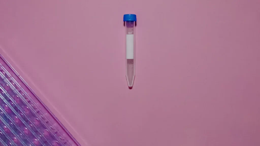
You start the western blot protocol by preparing your sample. You use a cell lysis buffer to break open cells and release proteins. If you skip protease inhibitors, proteins can degrade quickly. This leads to faint bands or no signal during protein analysis. Common pitfalls include unusual bands, high background, and patchy spots.
Tip: Always keep your samples cold and add inhibitors to prevent protein breakdown.
| Pitfall Type | Common Issues |
|---|---|
| High Background | Incomplete blocking, poor washing, too much antibody, low sample quality, membrane drying |
| Weak or No Signal | Incomplete transfer, unactivated membrane, wrong antibody, low antigen levels |
| Non-Specific Bands | Over-passaged cells, protein degradation, excessive sample loading |
| Smearing | Glycosylated proteins, lysate degradation |
| Patchy/Uneven Spots | Poor transfer, air bubbles, uneven agitation |
Gel Electrophoresis
You separate proteins by size using gel electrophoresis. If you use the wrong buffer or pH, bands may look distorted. Poor separation makes it hard to identify proteins. High-quality separation is key for reliable western blot results.
Protein Transfer
You move proteins from the gel to a membrane. You must control temperature because too much heat can damage proteins. Adjust voltage and time for efficient transfer. High molecular weight proteins need longer transfer times.
- Keep transfer settings optimized.
- Watch for air bubbles that block protein movement.
Blocking
You block the membrane to prevent non-specific binding. Non-fat dry milk, BSA, and normal serum work well. Tween 20 helps reduce background but must be washed out. Nitrocellulose membranes often lower background better than PVDF.
- Use protein-based blockers for most western blot protocols.
- Try polyvinylpyrrolidone for a hydrophilic barrier.
Antibody Incubation
You add primary and secondary antibodies to detect your protein. Antibody concentration affects specificity and sensitivity. Too much antibody increases background. Longer incubation can raise noise. Good blocking improves results.
- Use the right antibody concentration.
- Wash membranes well to remove weakly bound antibodies.
| Evidence Description | Impact on Western Blot Outcomes |
|---|---|
| Antibody concentration affects binding efficiency and specificity. | Higher concentrations increase binding but may cause non-specific interactions. |
| High antibody concentrations can lead to increased background noise. | Non-specific binding can overshadow specific signals. |
| Poor washing techniques exacerbate non-specific binding. | Inadequate washing increases background signal. |
Detection
You choose a detection method for protein detection. Chemiluminescent detection finds very low amounts of protein. Fluorescent detection offers a broad range and high sensitivity. Colorimetric detection has limited sensitivity.
| Detection Method | Sensitivity |
|---|---|
| Chemiluminescent | Very high (femtogram levels) |
| Fluorescent | High (comparable to chemiluminescence in NIR) |
| Colorimetric | Limited sensitivity |
Note: Always include controls like positive and negative lysates, loading controls, and no primary antibody controls to validate your western blot protocol.
Common Western Blot Issues

When you run a western blot, you may see several common problems. These issues can make it hard to get clear and reliable results. Knowing what to look for helps you fix mistakes early and improve your experiment.
High Background
You may notice a hazy or dark background on your membrane. This makes it hard to see your protein bands. High background happens often in western blot experiments. Many researchers face this problem. You might see it if you use too much antibody, skip proper blocking, or do not wash the membrane well.
- High background signal is a common problem in western blot experiments.
- Specific causes include insufficient blocking, high primary antibody concentration, and inadequate washing.
- Solutions involve reducing antibody concentration and increasing washing time.
Tip: Always check your blocking buffer and wash steps. Try lowering your antibody concentration if you see high background.
No Signal
Sometimes, you do not see any bands at all. This means your protein did not show up on the membrane. You might have lost your sample during transfer or used the wrong buffer. Problems with electrophoresis can also cause no signal. Make sure your sample is fresh and your detection method works.
Weak Signal
A faint band can mean your protein is present, but not enough to see clearly. You may have loaded too little sample or used a buffer that did not protect your protein. Weak signal can also happen if your antibody is too diluted. Check your sample concentration and buffer quality.
Non-Specific Bands
Extra bands that do not match your target protein can confuse your results. These bands often come from using too much antibody or loading too much sample. Sometimes, the buffer does not match the needs of your protein. You can fix this by adjusting your antibody and buffer.
Uneven Bands
Bands that look streaky or patchy can signal problems with electrophoresis or transfer. Air bubbles, uneven sample loading, or poor buffer mixing can cause this. Make sure you load your sample carefully and check your buffer before starting electrophoresis.
Troubleshooting High Background
Causes
High background in a western blot can make it hard to see your target bands. You may notice a cloudy or dark membrane. This problem often happens when you do not control each step of your protocol. Here are some common causes:
- You use too much antibody. High concentrations can stick to the membrane and create extra signal.
- You do not block the membrane well. Weak blocking lets the antibody bind where it should not.
- You skip or rush washing steps. Leftover antibody stays on the membrane and increases background.
- Your buffer does not have enough detergent, like Tween 20. This makes it harder to wash away unbound antibody.
- The membrane dries out during the process. Dry spots trap antibody and cause uneven background.
- You use milk-based buffer when detecting phosphorylated proteins. Milk can react with some antibodies and raise background.
- You forget to include protease inhibitors in your sample. Proteins break down and create unwanted bands.
- You do not prepare fresh lysates or keep them cold. Old or warm samples can degrade and cause extra signal.
- You load too much protein onto the gel. Overloading can make the membrane look messy.
- You do not check for known isoforms or post-translational modifications. These can appear as extra bands.
- You use the wrong gel percentage for your protein’s size. This can cause smearing and background.
- You skip positive and negative controls. Without controls, you cannot tell if the background comes from the sample or the protocol.
Tip: Always run a secondary antibody-only control. This helps you see if the secondary antibody binds non-specifically.
Solutions
You can lower high background in your western blot by making small changes to your protocol. Try these steps to get clearer results:
- Dilute your antibody more. Start with a higher dilution and adjust as needed.
- Increase the concentration of your blocking reagent. For example, use 7% non-fat dry milk instead of 5%.
- Block the membrane for a longer time or at a higher temperature. This gives better coverage.
- Add Tween 20 to your blocking buffer. This helps wash away unbound antibody.
- Include both blocking reagent and Tween 20 in your primary antibody dilution buffer.
- Wash the membrane longer and use more buffer. More washing removes extra antibody.
- Change your washing buffer to a stronger detergent, like SDS, if background stays high.
- Use non-fat dry milk as your blocking buffer. It often works better than BSA for lowering background.
- Always consult the recommended protocol for your antibody. Some antibodies need special buffers.
- Make sure the membrane stays wet during the whole process. Dry spots cause uneven background.
- Prepare fresh lysates for each experiment and keep them on ice. This keeps your protein stable.
- Always add protease inhibitors to your sample. This stops protein breakdown.
- Use pre-adsorbed secondary antibodies. These have less crossreactivity and lower background.
- Check for known isoforms and post-translational modifications of your protein. This helps you spot real bands.
- Adjust the gel percentage to match your protein’s size. This improves separation and reduces background.
- Include positive and negative controls in every western blot. Controls help you find the source of background.
| Step | Action to Reduce Background |
|---|---|
| Antibody | Use higher dilution, check protocol recommendations |
| Blocking | Increase concentration and time, use non-fat milk |
| Washing | Increase time and volume, use stronger detergent |
| Sample | Prepare fresh, keep cold, add inhibitors |
| Controls | Always include positive and negative controls |
Note: If you still see high background after these steps, try a secondary antibody-only control. This will show if the secondary antibody is causing the problem.
Troubleshooting No Signal
Causes
You may run a western blot and see no bands at all. This problem can frustrate you, but you can solve it by checking each step. Many issues can cause no signal. You might use too little protein in your sample. If you load less than 20–30 micrograms per lane, you may not detect your target. Sometimes, your protein degrades because you forget protease inhibitors. You need fresh samples to keep your protein intact.
Your antibody may not recognize the protein. Always check the datasheet or run a BLAST alignment to confirm specificity. If you use the wrong antibody concentration, you may miss the signal. Too little antibody gives weak or no bands. Too much antibody can cause ghost bands, which look hollow.
Problems with the buffer can also block detection. If you use sodium azide in your buffer, you can inhibit HRP activity. This stops the detection agent from working. You may also lose signal if you wash the membrane too much. Excessive washing can remove your protein from the membrane.
Transfer errors often cause no signal. If you do not set up the transfer correctly, your protein may stay in the gel. Air bubbles under the membrane block protein movement. You should always check the transfer with a reversible stain.
Here is a table that shows common causes and solutions for no signal in western blot experiments:
| Common Issues for No Signal in Western Blotting | Suggested Solutions |
|---|---|
| Antibody Concentrations Too Low | Use a higher concentration or incubate longer. |
| Antibody Concentrations Too High | Decrease concentrations if ghost/hollow bands appear. |
| Primary Antibody Does Not Recognize Antigen | Check datasheet or perform a BLAST alignment. |
| Detection Agent Inhibited | Avoid HRP with sodium azide or hemoglobin. |
| Protein is Undetectable | Load sufficient protein (~20-30 µg) and ensure it is not degraded. |
| Protein of Interest Not Abundant | Use enrichment steps or concentrate protein lysates. |
| Protein Did Not Transfer | Optimize transfer protocol by increasing time or voltage. |
Tip: Always include a positive control in your western blot. This helps you confirm that your protocol works and your sample contains the protein.
Solutions
You can fix no signal in your western blot by following a checklist. Start by checking your sample. Make sure you load enough protein. Aim for 20–30 micrograms per lane. Use fresh samples and add protease inhibitors to prevent degradation.
Check your buffer. Use sodium azide-free buffers to keep HRP active. If you use colorimetric or chemiluminescent detection, avoid hemoglobin in your buffer. Make sure your buffer matches the antibody and detection system.
Look at your antibody concentration. Titrate the antibody to find the best level. If you see no bands, try a higher concentration or longer incubation. If you see ghost bands, lower the concentration.
Review your transfer setup. Make sure you avoid air bubbles under the membrane. Use a reversible stain to check that your protein moved from the gel to the membrane. If you see no signal, increase the transfer time or voltage.
Reduce washing steps if you suspect protein loss. Too much washing can remove your protein from the membrane. Use gentle washing and check your protocol.
Here is a checklist to help you troubleshoot no signal in your western blot:
- Load enough protein in each lane (20–30 µg).
- Use fresh samples and add protease inhibitors.
- Use sodium azide-free buffer for HRP detection.
- Titrate antibody concentration for best results.
- Include a positive control to validate your experiment.
- Check transfer setup and avoid air bubbles.
- Use a reversible stain to confirm protein transfer.
- Reduce washing steps if you lose signal.
- Use the correct lysis buffer for your sample.
Note: If you still see no signal after these steps, check the antibody datasheet. Make sure your antibody matches your protein and species.
Troubleshooting Weak Signal
Causes
You may notice faint bands or almost invisible results during your western blot experiment. Weak signal often means you cannot see your target protein clearly. Several factors can lead to this problem. You might use too little sample, or your transfer step may not move enough protein onto the membrane. Deviations from the recommended protocol can cause low signal or high background, forcing you to repeat your experiment.
You may choose the wrong blocking buffer. Including Tween 20 during blocking can reduce antibody detection and weaken your signal. Using incorrect antibody dilution buffers also affects detection. Sometimes, excessive secondary antibody during antibody incubation leads to signal saturation, making it hard to interpret results. If you use chemiluminescent detection or fluorescent detection, improper buffer selection can lower sensitivity. Poor transfer efficiency or incomplete transfer can also cause weak bands. You may see this if you skip Ponceau S staining to check transfer quality.
- Using too little sample or protein
- Incomplete transfer of protein to the membrane
- Incorrect blocking buffer components (such as Tween 20)
- Wrong antibody dilution buffer
- Excessive secondary antibody during antibody incubation
- Poor detection method setup (chemiluminescent detection or fluorescent detection)
- Skipping transfer quality checks
Tip: Always check your transfer efficiency before moving to detection. This helps you catch problems early.
Solutions
You can improve weak signal in your western blot by making small changes to your protocol. Start by evaluating your transfer step. Use Ponceau S staining to confirm that protein has moved onto the membrane. If you see poor transfer, adjust your voltage or time. Try universal buffers instead of non-fat dry milk for blocking. Universal buffers help avoid masking antigens and can reduce blocking times. Alternatives like bovine serum albumin or PVP-40 work well as blocking agents. PVP-40 is nontoxic and biocompatible, making it a cost-effective choice for visualizing low-quality antibodies.
If you use chemiluminescent detection or fluorescent detection, make sure your buffer matches the detection system. The Antigen Retrieval for Western Blot Method (ARWB) can boost signal strength. Incubate your membrane in citrate buffer after transfer. Researchers found that this step increases band density and reveals bands that were previously invisible. This method is simple and economical.
- Check transfer efficiency with Ponceau S staining
- Use universal buffers for blocking
- Try PVP-40 or bovine serum albumin as blocking agents
- Match buffer to your detection method (chemiluminescent detection or fluorescent detection)
- Apply ARWB by incubating membrane in citrate buffer after transfer
Note: Always optimize your antibody incubation and detection steps for your specific protocol. Small adjustments can make a big difference in your western blot results.
Troubleshooting Non-Specific Bands
Causes
You may see extra bands on your western blot that do not match your target protein. These non-specific bands can confuse your results. Poor antibody quality often causes this problem. Some antibodies can bind to proteins that have similar sizes. For example, studies show that antibodies targeting amyloid precursor protein (APP) sometimes detect a 20 kDa band in human brain samples. This means you need to be careful when you analyze proteins with similar molecular weights.
Non-specific bands can also appear if you load too much protein onto the gel. When you use excessive protein, the membrane can become overloaded, and extra bands may show up. If you do not wash the membrane well, leftover antibodies can stick to the membrane and create unwanted bands.
- Poor antibody quality or specificity
- Excessive protein loading
- Insufficient washing during the western blot process
Tip: Always check your antibody datasheet and use recommended washing steps to reduce non-specific bands.
Solutions
You can reduce non-specific bands in your western blot by making a few changes to your protocol. Start by adjusting your antibody concentration. Try diluting your primary and secondary antibodies more. Lower concentrations help prevent unwanted binding. You should also load less protein into each well. This keeps the membrane from becoming overloaded.
Increase the concentration of your blocking agent. For example, use 7% non-fat dry milk instead of 5%. Block the membrane for a longer time or at a higher temperature. This helps cover spots where antibodies might stick. Use more detergent, like 0.05% Tween-20, in your wash buffer. This helps remove antibodies that do not bind specifically.
Here is a table with helpful modifications:
| Modification | Recommendation |
|---|---|
| Antibody Concentration | Dilute primary and secondary antibodies further |
| Protein Loading | Load less protein per well |
| Blocking Conditions | Increase blocker concentration and incubation time |
| Washing Techniques | Use more detergent in wash buffer and wash more thoroughly |
| Incubation Times | Shorten antibody incubation times |
You should also use positive and negative controls in every western blot. These controls help you spot non-specific bands and confirm your results.
Note: When you work with proteins of similar sizes, always analyze your bands carefully. Non-specific binding can happen even with good antibodies.
Troubleshooting Uneven Bands
Causes
You may see uneven bands when you run a western blot. This problem can make your results hard to read. Uneven bands often happen because of mistakes during the protocol. You need to watch each step closely to avoid this issue. Here are common causes you might encounter:
- Incomplete transfer of proteins from the gel to the membrane.
- Uneven sample loading onto the gel.
- Poor gel quality affecting protein migration.
- Inadequate blocking of the membrane leading to non-specific binding.
- Suboptimal antibody incubation conditions.
- Poor washing steps after antibody incubation.
- Inconsistent development of the chemiluminescent or chromogenic signal.
If you notice streaks or patchy bands, you should check your western blot setup. Problems like air bubbles or uneven agitation can also lead to these results.
Solutions
You can fix uneven bands in your western blot by making careful adjustments. Start by checking your equipment and reagents. Use the table below to match issues with solutions:
| Issue | Solution |
|---|---|
| High voltage during migration | Check the suggested voltage and decrease if necessary. |
| Gel too hot during migration | Run the gel at 4°C: on ice or in a cold room. |
| Uneven polymerization of gel | Check gel recipe for correct amount of TEMED and ensure gel is covered in buffer while setting. |
You should also follow these steps to improve your results:
- Check buffers for contamination and make fresh reagents if needed.
- Make sure the membrane is fully immersed during washes and antibody incubations.
- Gently remove any air bubbles, especially during transfer.
- Use a rocker or shaker for uniform agitation during incubations.
- Wash the electrophoresis unit thoroughly to remove protein or gel remnants.
- Filter conjugates to remove HRP aggregates.
- Reduce exposure time if bands appear too dark or streaky.
Tip: Always inspect your gel and membrane before starting your western blot. Small details can make a big difference in your results.
If you follow these steps, you can get clear and even bands in your western blot. Careful attention to each part of the protocol helps you avoid common mistakes.
Western Blotting Protocol Tips
Best Practices
You can improve your western blot results by following a set of protocol tips that experts recommend. These protocol tips help you avoid common mistakes and make your experiments more reliable. Here are some protocol tips you should use every time:
- Prepare your samples with the right lysis buffer. Test different buffers to find the one that works best for your protein. This protocol tip ensures you get the most protein out of your cells.
- Keep protein concentrations the same in every sample. This protocol tip helps you compare results between different experiments.
- Always add protease inhibitors during sample preparation. This protocol tip protects your protein from breaking down.
- Use SDS-PAGE gels that are uniform. Avoid high voltages and load the same amount of protein in each lane. This protocol tip gives you clear bands.
- Choose the right membrane. Use PVDF for proteins that are hard to detect and nitrocellulose for proteins that are easy to find. This protocol tip matches the membrane to your target.
- Titrate your antibodies. Adjust concentrations to find the best signal. This protocol tip saves you time and reagents.
- Try different blocking buffers. Avoid milk-based buffers if you study phosphorylated proteins. This protocol tip prevents unwanted background.
- Pick sensitive detection methods. Adjust film exposure times for the best results. This protocol tip helps you see even faint bands.
Following these protocol tips increases the reproducibility of your western blot and helps you avoid repeating experiments.
Quick Fixes
Sometimes you need protocol tips that give fast results. Use these quick protocol tips to fix problems right away:
| Protocol Tip | How It Helps |
|---|---|
| Use PVDF membrane | Improves signal for hard-to-detect proteins in your western blot. |
| Try a mix of blocking agents | Covers more sites on the membrane than BSA alone. |
| Heat at 50°C for 30 minutes | Boosts IgG detection in your western blot. |
| Acetone treat at 0°C, then heat | Gives the strongest signal in your western blot. |
| Add 0.5% glutaraldehyde | Increases sensitivity for small proteins. |
- Always check for air bubbles during transfer. This protocol tip prevents uneven bands.
- Use a rocker for even washing and incubation. This protocol tip ensures all parts of the membrane get treated the same.
- Filter your antibody solutions. This protocol tip removes clumps that can cause spots.
Try these protocol tips when you need a quick fix for your western blot. Small changes can make a big difference.
Mastering each step of your western blot protocol helps you solve problems quickly and get better results. When you use troubleshooting strategies, you improve the quality and reliability of your experiments. You can spot issues like air bubbles or nonspecific binding before they affect your data. Many researchers use trusted tools and reagents to support their work:
- Survey results on western blot instruments and reagents, including signal detection kits and troubleshooting issues
- Thermo Fisher SuperSignal West ECL reagents, often used in recent studies
Remember, troubleshooting is a normal part of science. Each challenge helps you learn and improve your skills.
FAQ
What should you do if you see no bands after running your western blot?
You should check your sample loading, antibody concentration, and transfer efficiency. Use a positive control to confirm your protocol works. Make sure you use fresh samples and add protease inhibitors.
How can you reduce high background on your membrane?
You can dilute your antibodies, increase washing steps, and use a stronger blocking buffer. Make sure your membrane stays wet. Try adding Tween 20 to your wash buffer for better results.
Why do you get non-specific bands in your western blot?
You may use too much antibody or load too much protein. Poor washing can also cause non-specific bands. Check your antibody datasheet and use recommended concentrations for best results.
What is the best way to check protein transfer from gel to membrane?
You can use Ponceau S staining to see if proteins moved onto the membrane. This quick test helps you spot transfer problems before detection.
Can you reuse membranes for western blotting?
You can reuse membranes after stripping them, but signal strength may decrease. Always follow the stripping protocol and check for leftover antibodies before reprobing.